Volumescope 2 SEM
3D SEM for large volume samples with serial block face imaging and multi energy deconvolution.
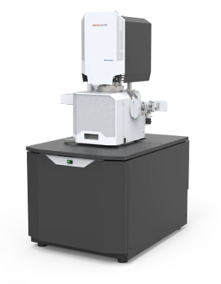 | The Thermo Scientific Volumescope 2 Scanning Electron Microscope (SEM) is our state-of-the-art serial block-face imaging system. Keep control of experiments with easy to use technology and protect a wide range of possible samples with tried and tested solutions for every acquisition step. Improving large-volume SEM by combining serial block-face imaging with multi-energy deconvolutionUnraveling the complex 3D architecture of cells and tissues in their natural context is crucial for structure-function correlation in biological systems and soft materials. Serial block-face SEM (SBF-SEM) combines in situ sectioning and imaging of plastic-embedded tissue blocks within the SEM vacuum chamber for automated, 3D reconstruction of large tissue volumes. Axial resolution was once limited by the minimum section thickness, but truly isotropic resolution is now possible with the addition of multi-energy deconvolution. Following in situ sectioning of the block-face using a diamond knife, the freshly exposed tissue is imaged several times using increasing accelerating voltages. These images are subsequently used in a deconvolution algorithm to derive several optical subsurface layers, forming a 3D subset. By repeating this cycle, the Volumescope 2 SEM offers isotropic datasets with 10 nm Z-resolution. | |
 Mouse brain sample imaged on the Volumescope 2 SEM. Fine features such as vesicles and synaptic clefts can be observed. Image courtesy of Dr. Gabor Nyiri, Institute of Experimental Medicine, Budapest, Hungary. |  Volume reconstruction of a mouse heart muscle. Data acquired by a combination of physical and virtual slicing in high vacuum mode. Volume = 16 x 15 x 11 μm3 with a nominal resolution of 5 x 5 x 10 nm. Specimen courtesy of Dr. Madesh Muniswamy, Temple University, USA. | |
Revealing the microstructure of polymers
High-resolution imaging in 3D is of great importance when trying to understand the microstructure and properties of polymer materials. The Volumescope 2 SEM combines in situ sectioning and imaging of polymers within the SEM vacuum chamber, in a fully automated fashion, for reconstruction of large volumes with truly isotropic 3D resolution. Visualizing a large volume at such resolutions is critical for revealing how regions with different microstructures affect the overall properties of the material. For example, the Volumescope 2 SEM can recreate a large representative volume of a filtration membrane, providing accurate transport properties that lead to enhanced performance predictions for new filter designs.
Key Features
Easy to use technologyKeep control of experiments with easy to use technology. Reuse jobs and system settings and select multiple regions of interest during job acquisition. Protect valuable samplesProtect valuable samples with tested solutions at every acquisition step: debris trap and swipe features ensure sample quality; low vacuum detector enables imaging of highly charged samples. | Visualize and navigate during acquisitionVisualize and navigate during acquisition with Thermo Scientific Amira Live Tracker Software to optimize/control your outcome/Automate large 3D volume acquisitions as well as reconstructions. Easy and fast mounting microtome exchangeEasy and fast mounting microtome exchange for normal SEM operation or automated array tomography with the addition of Thermo Scientific Maps Software. | |
For more details: https://www.thermofisher.com/tr/en/home/electron-microscopy/products/scanning-electron-microscopes/volumescope-2-sem.html